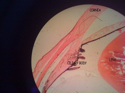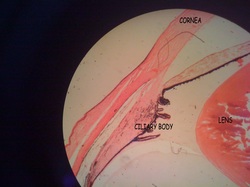
The posterior surface of the iris is covered by the retina. The inner layer of the retina, i.e. the layer facing the posterior chamber, is called the posterior epithelium of the iris. Both layers of the retina are pigmented, but pigmentation is heavier in the inner layer. In the region of the central opening of the iris, the pupil, the retina extends for a very short distance onto the anterior surface of the iris. The iridial stroma consists of a vascularized loose connective tissue rich in melanocytes in addition to macrophages and fibrocytes, which are all surrounded by a loose meshwork of fine collagen fibers. The anterior surface of the iris is not covered by an epithelium - instead of we find a condensation of fibrocytes and melanocytes, the anterior border layer of the iris.
The iris forms the aperture of the eye. Myoepithelial cells in the outer (or anterior) layer of the retina, i.e. the layer adjacent to the stroma of the iris, have radially oriented muscular extensions. These extensions form a flat sheet immediately beneath the anterior layer of the retina, the dilator pupillae muscle. Embedded in the central portion of the iridial stroma are smooth muscle cells which form the annular sphincter pupillae muscle. In humans, this muscle surrounds the pupil as a less than 1 mm wide and only 0.2 mm thick band. The two muscles regulate the size of the pupil.
Pupillary constriction, which is mediated by the sphincter pupillae muscle, is clinically refered to as miosis - dilation, mediated by the dilator pupillae muscle, as mydriasis.
The pigmentation of cells in the stroma and anterior border layer of the iris determines to color of the eyes. If cells are heavily pigmented the eyes appear brown. If pigmentation is low the eyes appear blue. Intermediate levels create shades of green and grey.
Reference : www.lab.anhb.uwa.edu.au
